Skip to main page content
U.S. flag

An official website of the United States government

Dot gov

The VSports app下载. gov means it’s official. Federal government websites often end in . gov or . mil. Before sharing sensitive information, make sure you’re on a federal government site. .

Https

The site is secure V体育官网. The https:// ensures that you are connecting to the official website and that any information you provide is encrypted and transmitted securely. .

Comparative Study
. 1999 Mar 16;96(6):2976-81.
doi: 10.1073/pnas.96.6.2976.

Functional differences between memory and naive CD8 T cells

Affiliations
Comparative Study

VSports - Functional differences between memory and naive CD8 T cells

B K Cho et al. Proc Natl Acad Sci U S A. .

Abstract

To determine how murine memory and naive T cells differ, we generated large numbers of long-lived memory CD8(+) T cells and compared them to naive cells expressing the same antigen-specific receptor (T cell receptor; TCR). Although both populations expressed similar levels of TCR and CD8, on antigen stimulation in vitro memory T cells down-regulated their TCR faster and more extensively and secreted IFN-gamma and IL-2 faster than naive T cells. Memory cells were also larger, and when freshly isolated from mice they contained perforin and killed target cells without having to be restimulated. They further differed from naive cells in requiring IL-15 for proliferation and in having a greater tendency to undergo apoptosis in vitro. On antigen stimulation in vivo, however, they proliferated more rapidly than naive cells. These findings suggest that, unlike naive T cells, CD8 memory T cells are intrinsically programmed to rapidly express their effector functions in vivo without having to undergo clonal expansion and differentiation VSports手机版. .


Figures
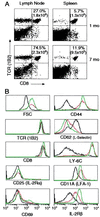
Figure 1
Figure 1
Persistence and surface phenotype of antigen-stimulated 2C cells. (A) Persistence. One and 7 months after immunization, lymph node and spleen cells of RAG1−/− recipients were stained with Abs to the 2C TCR and CD8. The percentages and total numbers (in parentheses) of 1B2+CD8+ T cells are shown. In the transferred naive cell population, 5–25% of the 1B2+ cells were CD8 and CD4; these cells persisted after the recipients were immunized. (B) Surface phenotype. Lymph node cells from naive 2C/RAG donors and immunized RAG1−/− recipients were stained with Abs to 2C TCR, CD8, and other cell-surface proteins. Cell size (forward light scatter, FSC) and expression of various markers were gated on live 1B2+CD8+ cells. Naive cells (black); memory cells at 1 month (red) and 7 months (green).
Figure 2
Figure 2
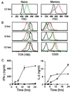
Memory cells respond more rapidly to antigen than naive cells. Purified memory and naive 2C cells were incubated with antigen, removed after 3, 6, and 12 hr, and stained with Abs to 2C TCR, CD8, and CD25. Forward light scatter (FSC) and expression of TCR and CD25 were gated on live CD8+ cells. No antigen added (black); memory cells plus antigen (red); naive cells plus antigen (green). (A) FSC. (B) Surface levels of TCR and CD25. (C) Secretion of IFN-γ and IL-2. Culture supernatants were assayed by ELISA for IFN-γ and IL-2. The symbols are as follows: memory cells with antigen (●) or without antigen (○); naive cells with antigen (■) or without antigen (□).
Figure 3
Figure 3
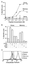
Memory 2C cells are cytolytic. (A) Comparison of cytolytic activities of memory and naive cells and 2C CTL clone. Purified cells were added at a 20:1 T cell to target cell ratio to 51Cr-labeled T2-Kb cells with various concentrations of SYRGL. 1B2 Ab to the 2C TCR was added at 60 μg/ml to verify that lysis was TCR dependent (open symbols). (B) The effect of various inhibitors on cytolytic activity of memory cells. Cytolytic assays were as in A, but before the addition of target cells and peptide, T cells were incubated with cycloheximide (CHX, 20 μg/ml) or concanamycin A (CMA, 2 mM) for 2.5 hr or with EGTA (4 mM) for 20 min. Inhibitors were present throughout the assay. (C) Memory cells express perforin. Memory cells, naive cells, and a 2C CTL clone (2C88) were stained with 1B2, fixed, permeabilized, and stained intracellularly with an anti-perforin Ab (P1–8). Intracellular perforin of 1B2+ cells is shown. Control refers to memory cells stained only with the secondary Ab. Peak 1, control; peak 2, naive cells; peak 3, memory cells; and peak 4, CTL clone 2C88.
Figure 4
Figure 4
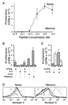
Proliferation of memory and naive cells in vitro. (A) Comparison of the proliferation of purified memory and naive cells by [3H]thymidine incorporation. (B and C) Proliferation under different conditions. Cells were stimulated with SYRGL in the presence or absence of IL-2 (100 units/ml) or human recombinant IL-15 (100 ng/ml), or with Con A (2 μg/ml) or PMA plus ionomycin (25 ng/ml and 0.5 μM, respectively). Naive cells (N) are shown in hatched columns and memory cells (M) in open columns. (D) Apoptosis of memory cells following antigen stimulation. Memory cells were incubated with SYRGL and irradiated B6 splenocytes as in Fig. 2. After 3-hr, cells were stained with Abs to the 2C TCR, CD8, and with Annexin V. The Annexin V expression was gated on 1B2+CD8+, propidium iodide-negative cells. Peak 1, unstimulated cells at time zero; peak 2, unstimulated cells at 3 hr; and peak 3, antigen-stimulated cells at 3 hr.
Figure 5
Figure 5
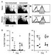
Proliferation of memory T cells in vivo. (A) Memory or naive cells were transferred into nonirradiated B6 recipients and immunized 3 days later. Mice were injected i.p. with BrdUrd 5 hr before immunization and were maintained on BrdUrd in drinking water. Three and 5 days following immunization, cells from draining lymph nodes (axial, lateral, and inguinal) were stained with Abs to TCR, CD8, and BrdUrd to determine the number and percentage of 1B2+CD8+ cells that incorporated BrdUrd (see arrow). Specificity of the BrdUrd stain is shown by its inhibition by 0.1 mM soluble BrdUrd (+sBrdUrd). The plots shown are for recipients on day 5 after immunization. (B) Percentages of 2C CD8+ cells (Left) and total numbers (Right) in lymph nodes 3 and 5 days after immunization. The symbols are as follows: naive recipients with immunization (□) or without immunization (■); memory recipients with immunization (○) or without immunization (●). Each symbol represents an individual mouse. Horizontal lines are mean values.

References

    1. Zinkernagel R M, Bachmann M F, Kündig T M, Oehen S, Pircher H, Hengartner H. Annu Rev Immunol. 1996;14:333–367. - PubMed
    1. Ahmed R, Gray D. Science. 1996;272:54–60. - PubMed
    1. Sprent J. Curr Opin Immunol. 1997;9:371–379. - PubMed
    1. Eisen H N. Harvey Lect. 1966;60:1–34. - PubMed
    1. Steiner L A, Eisen H N. J Exp Med. 1967;126:1185–1205. - PMC - PubMed

Publication types

MeSH terms