

Abstract
Arecoline, the main alkaloid of areca nut, is well known for its role in inducing submucosal fibrosis and oral squamous cell carcinoma (OSCC), however the mechanism remains unclear V体育官网入口. The aim of this study was to establish an arecoline‐induced epithelial‐mesenchymal transformation (EMT) model of OSCC cells and to investigate the underlying mechanisms. CAL33 and UM2 cells were induced with arecoline to establish an EMT cell model and perform RNA‐sequence screening. Luminex multiplex cytokine assays, western blot, and RT‐qPCR were used to investigate the EMT mechanism. Arecoline at a concentration of 160 μg/ml was used to induce EMT in OSCC cells, which was confirmed using morphological analysis, transwell assays, and EMT marker detection. RNA‐sequence screening and Luminex multiplex cytokine assays showed that many inflammatory cytokines (such as serum amyloid A1 [SAA1], interleukin [IL]‐6, IL‐36G, chemokine [CCL]2, and CCL20) were significantly altered during arecoline‐induced EMT. Of these cytokines, SAA1 was the most highly upregulated. SAA1 overexpression induced EMT and promoted the migration and invasion of CAL33 cells, while SAA1 knockdown attenuated arecoline‐induced EMT. Moreover, arecoline enhanced cervical lymph node metastasis in an orthotopic xenograft model of the tongue established using BALB/c nude mice. Our findings revealed that arecoline induced EMT and enhanced the metastatic capability of OSCC by the regulation of inflammatory cytokine secretion, especially that of SAA1. Our study provides a basis for understanding the mechanism of OSCC metastasis and suggests possible therapeutic targets to prevent the occurrence and development of OSCC associated with areca nut chewing.
Keywords: arecoline, epithelial‐mesenchymal transition, inflammatory cytokines, oral squamous cell carcinoma, SAA1
Our findings uncovered that arecoline induced EMT and enhanced the metastatic capability of OSCC via regulation of inflammatory secretion, especially the secretion of SAA1. Our study provided a basis for understanding the mechanism of OSCC metastasis and a possible therapeutic target to prevent the occurrence and development of OSCC associated to the areca nut chewing V体育2025版.
![]()
"V体育安卓版" Abbreviations
- CDX
cell‐derived xenograft
- CL
cell lysate
- CS
cell supernatant
- EMT
epithelial‐mesenchymal transition
- LN
lymph node
- NF‐κB
nuclear factor kappa B
- OSCC
oral squamous cell carcinoma
- SAA1
serum amyloid A1
- SsnB
sparstolonin B
1. INTRODUCTION
The incidence of OSCC is increasing, and it is now the leading cause of cancer‐related death, mainly due to the high metastasis and recurrence rates VSports. 1 Smoking, alcohol, areca nut chewing, and human papillomavirus (HPV) infection are common risk factors for the development of OSCC worldwide. 2 , 3 , 4 In Western countries, the major risk factors for oral cancer are cigarette smoking, alcohol consumption, and HPV infection, whereas habitual chewing of areca nuts is considered an important risk factor for oral cancer and oral mucosal diseases in South and Southeast Asian countries. 5 , 6 .
Wang H et al reported that zinc finger protein 703 (ZNF703) can regulate the cell cycle and EMT by activating the PI3K/AKT/GSK‐3β signaling pathway in OSCC associated with areca nut VSports app下载. 7 Areca nut extracts can induce the generation of reactive oxygen species (ROS), therefore causing further autophagy and promoting OSCC metastasis. 8 Some studies also reported that areca nuts trigger cellular inflammatory responses and promote tumor progression through the regulation of several immune mediators, including IL‐6, TGF‐β, EGFR, ERK, and Ras. 9 , 10 , 11 Each component in areca nut extracts may individually, synergistically, and in a coordinated manner contribute to the occurrence or progression of OSCC. 5 Despite these advancements, the exact mechanisms underlying the carcinogenic and metastasis‐promoting effects of areca nut remain unclear.
Arecoline, the major alkaloid in areca nut, is considered the most significant carcinogen. 12 Previous studies have suggested that arecoline can increase the methylation levels of dual‐specificity protein kinase phosphatase 4 (DUSP4) 13 and sirtuin 1 (SIRT1), 6 leading to tumorigenesis and tumor metastasis in OSCC. EMT is considered to be a vital and fundamental process in the progression and metastasis of OSCC. A study in HK2 cells showed that arecoline treatment upregulated the expression of phosphorylated ERK to induce EMT and promoted renal fibrosis. 14 Another study reported that Slug expression contributes to myofibroblast transdifferentiation. 15 Therefore, studies are necessary to reveal the mechanisms by which arecoline contributes to OSCC progression and metastasis V体育官网.
In the present study, we hypothesized that arecoline promotes OSCC metastasis through EMT by increasing the secretion of various inflammatory cytokines. We successfully established an arecoline‐induced OSCC cell model with EMT phenotypic and molecular characteristics. We found using RNA‐sequence screening and Luminex multiplex cytokine assays that multiple inflammatory cytokines, such as serum amyloid A1 (SAA1), interleukin (IL)‐6, IL‐36G, and chemokine (CCL)2, were upregulated in arecoline‐induced OSCC cells. Of these cytokines, SAA1 was the most highly upregulated. Moreover, a series of in vitro and in vivo experiments demonstrated that arecoline treatment dramatically promoted metastasis of OSCC through regulation of the SAA1 pathway. This study suggested that SAA1 might be a potential therapeutic target for areca nut‐associated OSCC.
"VSports注册入口" 2. MATERIALS AND METHODS
2.1. Cell culture and treatment
Human OSCC cell lines CAL33 and UM2 were cultured in DMEM supplemented with 10% FBS, penicillin (100 U/mL), and streptomycin (100 μg/mL) (Invitrogen, Carlsbad, CA, USA). All cell lines were maintained in a standard humidified incubator with 5% CO2 at 37℃.
CAL33 and UM2 cell lines were treated with arecoline (Sigma, Carlsbad, CA, USA) to establish the EMT model of OSCC. When cell confluency reached approximately 80%, OSCC cells were initially treated with half of the IC50 dose (80 μg/mL) of arecoline, and then the cells were passaged. Subsequently, 20 μg/mL of arecoline were gradually added to the culture medium to reach concentrations ranging from 80 to 160 μg/mL for c. 20 d until the OSCC cell morphology changed from a polygon to a stable spindle shape. Arecoline concentration was maintained at 160 μg/mL for the following experiments.
Recombinant human SAA1 (Catalog #C633‐10, NovoProtein systems) was used at a concentration of 200 ng/mL. TLR2/4 inhibitor sparstolonin B (SsnB) (Catalog #1259330‐61‐4, Sigma‐Aldrich) was used at 20 μM. siRNAs were purchased from RiboBio (Guangzhou, China). The siRNA was transfected into cells using Lipofectamine 3000 (L3000015, Invitrogen) in accordance with the manufacturer's instructions. The SAA1‐siRNA1 and SAA1‐siRNA2 sequences were 5′‐GATCAGGCTGCCAATGAAT‐3′ and 5′‐GAGAGAATATCCAGAGATT‐3′, respectively.
2.2. Invasion and migration assays
CAL33 cells treated with arecoline, or not, at a density of 6 × 10⁴ cells/well in 200 μL serum‐free medium were seeded into the upper chamber of 24‐well transwell chambers (8 μm pore, costar) coated without (migration) and with (invasion) Matrigel (BD Biosciences, USA). The lower chambers were filled with 500 μL of medium containing 10% FBS. After 24 h (migration) or 48 h (invasion) of incubation, cells on the lower surface of the inserts were stained with 0.1% crystal violet. Five images per chamber were captured using an inverted microscope under ×100 magnification (Axio, ZEISS, Germany), and the number of cells was counted using ImageJ software.
2.3. RNA‐sequencing (RNA‐seq) and data processing
Total RNA was extracted from cells using RNAzol reagent (Molecular Research Center, Inc, USA). RNA integrity was assessed using the RNA Nano 6000 Assay Kit for the Bioanalyzer 2100 system (Agilent Technologies, CA, USA). Transcriptome sequencing was performed using next‐generation technology. After cluster generation, the library preparations were sequenced on an Illumina Novaseq platform. Differential expression analysis of 2 conditions/groups (2 biological replicates per condition) was performed using the DESeq2 R package (1.16.1). DESeq2 provides statistical routines for determining differential expression in digital gene expression data using a model based on a negative binomial distribution. The resulting P values were adjusted using the Benjamini–Hochberg approach for controlling the false discovery rate. Genes with an adjusted P value < .05 found using DESeq2 were assigned as differentially expressed. Gene Ontology (GO) enrichment analysis was implemented using the clusterprofiler R package. The clusterprofiler R package was used to test the statistical enrichment of differentially expressed genes in the Kyoto Encyclopedia of Genes and Genomes (KEGG) pathways (http://www.genome.jp/kegg/).
2.4. Real‐time quantitative PCR (RT‐qPCR)
Total RNA was extracted from cells using RNAzol reagent (Molecular Research Center, Inc). Reverse transcription was performed using PrimeScript RT reagent kit in accordance with the manufacturer's instructions (TaKaRa, Japan). RT‐qPCR was performed using the SYBR PrimeScript RT‐PCR kit (Roche, USA) following the manufacturer's protocol. The experiment was performed in triplicate. The relative expression levels of mRNA were computed using the 2−ΔΔCt method, where GAPDH was used as an internal control. The primers used in this study are listed in Table S1.
2.5. Western blotting
Cells were harvested and lysed in radioimmunoprecipitation buffer (Beyotime, China). The protein concentration was measured using a bicinchoninic acid (BCA) protein assay kit (CWBIO, China). Equal amounts of protein were separated using 10% SDS‐PAGE and then transferred to polyvinylidene fluoride (PVDF) membranes (Millipore, USA). The membranes were blocked with 2% skimmed milk or BSA for 2 h and incubated with primary antibodies at 4°C overnight and then incubated with secondary antibodies (EMAR, China) for 1 h at room temperature. The signal on the membrane was detected using the ECL reagent (ECL Ultra Western HRP Substrate, Millipore) and the GeneGnome XRQ system (Syngene, USA). Primary antibodies included those against SAA1 (ab207445‐100, Abcam, UK), GAPDH, Snail, E‐cadherin, N‐cadherin, and vimentin (Cell Signaling Technology, Danvers, MA, USA); GAPDH was used as the internal control. The ratio of the gray value of a target band to the gray value of the GAPDH band indicated the relative target protein expression.
2.6. Luminex multiplex cytokine assays
The cell culture supernatants and lysates were collected and utilized to carry out human Luminex multiplex cytokine assays (R&D system, USA) following the manufacturer's protocol. Briefly, the sample was added to a mixture of color‐coded beads pre‐coated with analyte‐specific capture antibodies. Biotinylated detection antibodies specific to the analytes of interest were then added to form an antibody‐antigen sandwich. Beads were read on the dual‐laser flow‐based detection instrument Luminex 200™ or FlexMap® analyzer. One laser classifies the bead and determines the analyte that is being detected. In addition to flow‐based analyzers, magnetic beads were used, and the analysis was performed using the Luminex MAGPIX® Analyzer. A magnet in the MAGPIX analyzer captures and holds the magnetic beads in a monolayer, while 2 spectrally distinct light‐emitting diodes (LEDs) illuminate the beads. One LED identifies the analyte that is being detected, and the second LED determines the magnitude of the PE‐derived signal. Each well was imaged with a charge coupled device (CCD) camera.
"VSports手机版" 2.7. Orthotopic xenograft model
All animal experiments were approved by the Animal Research Ethics Committee of Sun Yat‐Sen University (2 020 000 303). All BALB/c nude mice (weight 18‐22 g, male) were purchased from the animal facility of Sun Yat‐Sen University and housed in a specific pathogen‐free environment and treated in accordance with the guidelines of the Institutional Animal Use and Care Committee. First, CAL33 cells were transfected with the luciferase gene (GeneCopoeia Inc, USA). Approximately 0.6 million CAL33 cells suspended in 50 μL PBS in the presence or absence of arecoline were injected into the side of the tongue to investigate the effect of arecoline on the tumorigenesis and metastasis of OSCC in vivo. Tumor size and cervical LN metastasis of OSCC were observed at 1 wk, 2 wk, and 3 wk. After 3 wk, mice were sacrificed. The cervical LNs were isolated for H&E staining. For in vivo scans, mice were anesthetized using a mixture of oxygen/isoflurane inhalation and positioned with legs fully extended. The fluorescence images of mice were acquired using an In Vitro Imaging System (IVIS) Spectrum (PerkinElmer, Waltham, MA, USA). Furthermore, immunohistochemical staining was used to detect the expression of SAA1, cytokeratin, and HLA class I in the xenograft tissues or cervical LN as previously described. 16 The primary antibodies were SAA1 (Catalog #ab207445, Abcam, 1:1000), pan‐cytokeratin (Catalog #bs1712R, Bioss, 1:200) and HLA class I (Catalog #15240‐I‐AP, proteintech, 1:200).
2.8. Statistical analysis (VSports最新版本)
Bar graphs were plotted using GraphPad Prism 8 software (GraphPad, San Diego, CA, USA), and all data were expressed as the mean ± standard deviation (SD). Unless otherwise indicated, comparisons between 2 groups were performed using an unpaired, 2‐tailed t test. A P value of less than .05 was considered statistically significant.
3. RESULTS
3.1. Arecoline induced EMT and enhanced migration and invasion of OSCC cells (V体育ios版)
To verify whether arecoline could induce EMT in OSCC cells, CAL33, and UM2 cells were exposed to arecoline as described above. Consequently, the arecoline‐induced EMT model of OSCC cells was successfully established. As shown in Figure 1A,B, we observed under an optical microscope that CAL33 and UM2 cells had a polygonal shape in the control medium and acquired a spindle‐like shape following arecoline exposure. Furthermore, compared with control cells, migration (Figure 1C,D) and invasion (Figure 1E,F) were enhanced in arecoline‐treated OSCC cells. Additionally, RT‐PCR and western blot analyses (Figure 1G,H) also showed that the expression of E‐cadherin was decreased and that of N‐cadherin, vimentin, and Snail was increased in arecoline‐treated CAL33 and UM2 cells compared with that in untreated cells. Taken together, these results suggested that arecoline induces EMT and promotes migration and invasion of OSCC cells.
FIGURE 1.
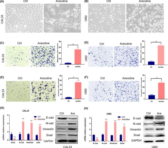
Arecoline‐induced EMT and enhanced migration and invasion of OSCC cells. A, B, OSCC cells lost cell‐cell adhesion and their morphology changed from a polygon shape to a spindle‐like shape after treatment with arecoline. OSCC cells enhanced migration (C, D) and invasion (E, F) capabilities after treatment with arecoline. G, H, RT‐qPCR and western blots were performed to detect the expression of EMT markers such as E‐cad, N‐cad, Snail, and vimentin. The expression levels of N‐cad, Snail, and vimentin were upregulated and E‐cad was downregulated. Data are shown as mean ± SD of 3 independent experiments. Are, Arecoline. *P value < .05, **P value < .01, ***P value < .001
3.2. Arecoline regulated global mRNA expression during EMT of OSCC
To further investigate the mechanism by which arecoline induces EMT, the transcriptome changes in arecoline‐exposed cells were assessed using RNA‐seq analysis. In total, the microarray chips detected 21 754 genes (Figure 2A). The results revealed 748 upregulated genes and 595 downregulated genes between arecoline‐treated OSCC cells and control cells. The top 20 upregulated and downregulated genes are shown in Table 1 and Table 2, respectively. GO analysis showed that the differentially expressed genes were significantly associated with important oncogenic pathways, including inflammatory response, positive regulation of cell migration, and extracellular matrix (Figure 2B). KEGG and Reactome enrichment analysis showed that these genes were associated with important cancer‐related functions, including cytokine‐cytokine receptor interaction, focal adhesion, ECM receptor interaction, and PI3K‐Akt signaling pathways (Figures 2C and S1A,B). Gene Set Enrichment Analysis (GSEA) showed that the upregulated genes were closely associated with chemokine signaling, cell adhesion, the ECM receptor signaling pathway, and the NF‐κB signaling pathway (Figures 2D,E and S1C). These results indicated that the inflammatory response and pro‐inflammatory cytokine secretion might be associated with OSCC related to arecoline.
TABLE 1.
Top 20 upregulated genes from RNA sequencing
| Gene name | Log2 fold change | P value |
|---|---|---|
| SAA1 | 14.13064681 | 2.41E‐45 |
| S100A7 | 13.6451131 | 9.29E‐42 |
| SAA2 | 11.65514997 | 1.81E‐27 |
| CCL20 | 11.05594764 | 2.28E‐45 |
| TNIP3 | 10.674796 | 6.20E‐21 |
| IL6 | 10.45255066 | 1.55E‐19 |
| S100A7A | 10.32225141 | 9.85E‐19 |
| CCL2 | 10.25240534 | 2.80E‐18 |
| IL36G | 10.14636727 | 1.27E‐17 |
| CSF3 | 9.572997768 | 2.38E‐14 |
| IL32 | 9.556531417 | 2.89E‐14 |
| MSC | 9.361792709 | 3.28E‐13 |
| CES1P1 | 9.222612091 | 1.52E‐12 |
| CSF2 | 8.842736918 | 9.92E‐11 |
| KRT75 | 8.815316609 | 1.70E‐10 |
| CES1 | 8.669806402 | 6.95E‐10 |
| RELN | 8.638856514 | 9.30E‐10 |
| CHI3L1 | 8.43776544 | 5.75E‐09 |
| S100A8 | 8.257737426 | 3.69E‐44 |
| IGFBP3 | 8.225816784 | 8.16E‐35 |
TABLE 2.
Top 20 downregulated genes from RNA sequencing
| Gene name | Log2 fold change | P value |
|---|---|---|
| DACH1 | −10.0776003 | 2.59E17 |
| SCGB1A1 | −9.238595727 | 9.72E13 |
| N4BP2L1 | −8.627927345 | 6.95E10 |
| AL590434.1 | −8.544081871 | 1.68E09 |
| CHST9 | −8.5150144 | 2.27E09 |
| VPS37D | −8.360184249 | 1.09E08 |
| SPINK8 | −8.149373205 | 5.79E08 |
| LGALS8‐AS1 | −7.902398107 | 5.06E07 |
| SOSTDC1 | −7.85678892 | 7.40E07 |
| EGFL6 | −7.604228688 | 5.45E06 |
| TLR1 | −7.547941704 | 5.45E06 |
| CASKIN1 | −7.547941704 | 5.45E06 |
| CFL1P1 | −7.489369202 | 8.28E06 |
| CHAD | −7.364568024 | 1.96E05 |
| FGF13 | −7.297870637 | 3.05E05 |
| HESX1 | −7.22793966 | 4.78E05 |
| ACTG1P1 | −7.077005484 | 0.00012 |
| LINC02028 | −7.077005484 | 0.00012 |
| AP002992.1 | −7.077005484 | 0.00012 |
| HS3ST6 | −6.99517173 | 0.000194 |
FIGURE 2.
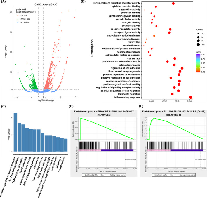
Arecoline regulated global mRNA changes in OSCC cells. A, Volcano plot depicts upregulated and downregulated genes in CAL33‐Arecoline (CAL33_A) vs CAL33‐Control (CAL33_C) (log2 fold change > 1.0, P value adjusted (padj) < .05). B, Bubble plots of GO analysis showed that upregulated genes were predominantly enriched in the inflammatory response signaling pathway. C, KEGG pathway enrichment analysis of transcriptome sequencing revealed that signaling pathway changes were mainly enriched in the cytokine‐cytokine receptor pathway, and PI3K‐Akt signaling pathway. GSEA of differentially expressed genes might be enriched in chemokine signaling pathway (D) and cell adhesion molecules (E)
VSports - 3.3. Arecoline enhanced secretion of pro‐inflammatory cytokines in OSCC cells
To understand the role of pro‐inflammatory cytokines in OSCC, we analyzed the signature profiles of the IL family, CCL family, tumor necrosis factor (TNF) superfamily, colony‐stimulating factor (CSF) family, and acute‐phase proteins (APPs) using publicly available datasets of oral tumor samples. We analyzed the database on The Cancer Genome Atlas (TCGA) and found that IL1A, IL32, and IL36G (Figure S2A) were highly expressed in OSCC. Both CCL2 and CCL20 were also highly expressed (Figure S2B). In addition, the expression levels of TNF‐and CSF were highly increased (Figure S2C,D). However, the expression of SAA1/2 was not significantly increased (Figure S2E). The results from TCGA also showed that the expression of some inflammatory factors was closely related to the prognosis and various clinicopathological parameters of OSCC patients. OSCC patients with elevated IL1A, CCL20, and CSF2 expression levels had a significantly worse prognosis (Figure S2F). In addition, high CCL20 expression was also positively correlated with the clinical stage of OSCC, but not with the T and N stages (Figure S2G). Taken together, these results indicated that pro‐inflammatory factors in OSCC may serve as molecular drivers for OSCC aggressiveness.
Luminex multiplex cytokine assays and RT‐qPCR were performed to determine whether the arecoline‐induced EMT of OSCC cells was mediated by inflammatory factors. The cytokine array results showed that the secretion of SAA1, CCL2, S100A8, IL‐6, and IL‐36G was significantly upregulated but that of CCL20 was downregulated in arecoline‐treated OSCC cells (Figure 3A,B). Consistent with the cytokine array results, arecoline treatment significantly increased mRNA expression levels of SAA1, CCL2, S100A8, IL‐6, and IL‐36G, and decreased CCL20 expression using RT‐qPCR analysis (Figure 3C).
FIGURE 3.
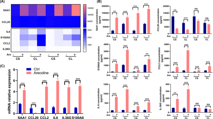
Arecoline enhanced secretion of pro‐inflammatory cytokines in OSCC cells. A, Cytokine profiles of CS and CL from CAL33 cells treated with or without arecoline were screened using the Luminex multiplex cytokine assays. B, Most cytokines, such as SAA1, CCL2, S100A8, IL‐6 and IL‐36G, were significantly increased, while CCL20 was decreased. C, mRNA levels of 6 pro‐inflammatory cytokines in OSCC cells were measured using RT‐qPCR. Data are shown as mean ± SD of 3 independent experiments. *P value < .05, **P value < .01, ***P value < .001
3.4. SAA1‐induced EMT of OSCC cells
Notably, SAA1 showed the highest fold change in arecoline‐treated cells compared with control cells (Table 1). Therefore, we next investigated whether SAA1 plays a role in cell migration and invasion. Cells were treated with 200 ng/mL of human recombinant SAA1. Following treatment with SAA1 the morphology of CAL33 and UM2 cells changed from a polygon to a fusiform‐like form (Figure 4A,B). Consistently, SAA1 treatment enhanced migration (Figure 4C,D) and invasion (Figure 4E,F) of OSCC cells. In addition, RT‐qPCR and western blot analysis showed that SAA1 treatment notably increased the expression of the mesenchymal markers N‐cadherin, vimentin, and Snail and reduced that of the epithelial marker E‐cadherin (Figure 4G,H) in OSCC cells. These findings indicated that SAA1 promotes the migration and invasion of OSCC cells by EMT.
FIGURE 4.
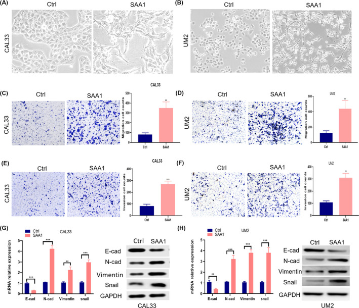
SAA1 promoted migration and invasion of OSCC cells. A, B, The morphology of OSCC cells changed from a polygon shape to a spindle‐like shape and cells lost cell‐cell adhesion after treatment with recombinant SAA1. OSCC cells were enhanced in migration (C, D) and invasion (E, F) after treated by recombinant SAA1. G, H, RT‐qPCR and western blot were used to detect the expression of EMT‐associated molecules. Expression levels of N‐cad, Snail, and vimentin were increased, while E‐cad was inhibited. Data are shown as mean ± SD of 3 independent experiments. *P value < .05, **P value < .01, ***P value < .001
Moreover, TLR2/4 inhibitor sparstolonin B (SsnB) was used to confirm whether TLR2/4, one of SAA1 receptor, played a role in the SAA1‐induced EMT through arecoline. As shown in Figure 5A,B, SsnB treatment hindered the morphological changes in CAL33 and UM2 cells from a polygon to a fusiform‐like shape. Consistently, SsnB treatment decreased the migration (Figure 5C,D) and invasion (Figure 5E,F) of OSCC cells. In addition, RT‐qPCR and western blot analysis showed that SsnB treatment notably decreased the expression levels of the mesenchymal markers N‐cadherin, vimentin, and Snail and increased those of the epithelial marker E‐cadherin in OSCC cells (Figure 5G,H).
FIGURE 5.
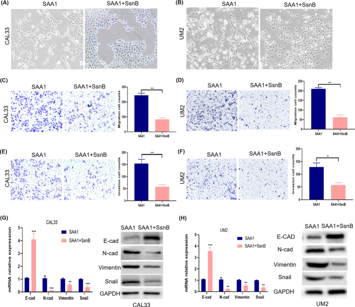
SAA1 promoted migration and invasion of OSCC cells were inhibited by TLR2/4 inhibitor. A, B, The morphology of OSCC cells changed from a spindle‐like shape to a polygon shape when TLR2/4 inhibitor sparstolonin B (SsnB) was used. The capabilities of OSCC cells were attenuated in migration (C, D) and invasion (E, F) after treatment with recombinant SAA1 combined with SsnB. G, H, RT‐qPCR and western blots were used to detect the expression of EMT‐associated molecules. Expression levels of N‐cad, Snail, and vimentin were inhibited, while E‐cad was elevated in OSCC cells treated with SAA1 combined with SsnB. Data are shown as mean ± SD of 3 independent experiments. *P value < .05, **P value < .01, ***P value < .001
3.5. SAA1 was critical for arecoline‐induced EMT in OSCC cells
To further determine whether SAA1 was involved in arecoline‐induced EMT, siRNA was used to suppress the expression of SAA1 in OSCC cells. We knocked down SAA1 in CAL33 and UM2 cell lines with siRNA and generated the siRNA‐SAA1 CAL33 and UM2 cell lines, as well as the corresponding control cell lines. SAA1 mRNA and protein levels were determined in these cell lines (Figure 6A). The morphology of CAL33 and UM2 cells did not change significantly after SAA1 knockdown, however the fusiform morphology (Figure S3) was visibly reversed in arecoline‐treated OSCC cells with SAA1 knockdown. The migration and invasion capabilities of OSCC cells were inhibited after SAA1 was knocked down in OSCC cells (Figures 6B,C and S4). Additionally, the enhanced migration and invasion capabilities in the arecoline‐induced OSCC cells were abolished after SAA1 knockdown (Figures 6D,E and S4). Furthermore, the arecoline‐induced changes in EMT biomarkers in OSCC cells were reversed by SAA1 knockdown (Figure 6F,H). Collectively, these results demonstrated that SAA1 is essential for arecoline‐induced EMT of OSCC cells.
FIGURE 6.
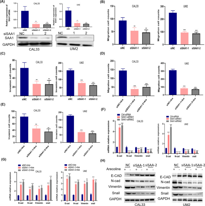
SAA1 knockdown inhibited arecoline‐induced EMT in OSCC cells. RT‐qPCR and western blot were used to verify the expression of SAA1 in SAA1‐knockdown CAL33 and UM2 cells. B, C, Effect of cell migration (B) and invasion (C) in OSCC cells after SAA1 knockdown. D, E, Effect of cell migration (D) and invasion (E) in arecoline‐exposed OSCC cells treated with or without SAA1 knockdown. F, G, Relative mRNA expression levels of EMT‐associated molecules in OSCC cells after SAA1 knockdown (F), or in arecoline‐exposed OSCC cells treated with or without SAA1 knockdown (G). H, Protein expression levels of EMT‐associated molecules in OSCC cells or arecoline‐exposed OSCC cells. Data are shown as mean ± SD of 3 independent experiments. *P value < .05, **P value < .01, ***P value < .001. Are, Arecoline
3.6. Arecoline promoted the metastasis of OSCC in vivo (V体育ios版)
To further explore the role of arecoline in OSCC in vivo, CAL33 cells treated or not treated with arecoline were labeled with luciferase and injected into the tongue tissues of nude mice. The orthotopic tumorigenesis of the tongue and cervical LN metastasis was observed after 3 wk. The bioluminescence signal of mice was higher in the arecoline group than in the control group (Figure 7A). No obvious difference in tumor size of the tongue tissues between the control and arecoline groups was found, however cervical LN metastatic tumors were found (Figures 7B and S5). Furthermore, tumors formed in the arecoline‐induced group displayed higher SAA1 levels than those in the control group (Figure 7C). Immunohistochemistry showed that cytokeratin and HLA class I were strongly expressed in cervical LN metastatic tumors (Figure 7D). Histological results showed that 4 out of 6 mice in the arecoline group and 1 out of 6 mice in the control group developed LN metastasis (Figure S5). The rate of cervical LN metastasis in the arecoline group increased by 50% compared with that in the control group. These results suggested that arecoline promotes the metastasis of CAL33 cells in vivo.
FIGURE 7.
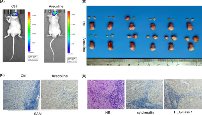
Arecoline promoted metastasis of OSCC in vivo. CAL33 cells treated with or without arecoline were inoculated into the tongues of nude mice. A, Luminescence was observed in the tongue and cervical LNs using an in vivo imaging system (IVIS) at 3 wk after cell injection. B, Representative photographs of tongue tumors and cervical LNs were shown. C, Expression of SAA1 in metastatic lymph nodules was examined by IHC. D, Representative immunohistochemical images showing the expression of cytokeratin and HLA class I in metastatic lymph nodules
4. DISCUSSION (VSports app下载)
Extensive epidemiologic evidence has demonstrated that OSCC is more prevalent in Southeast Asian than in Western countries, probably because Southeast Asians have the common habit of chewing areca nut, which increases the risk of OSCC. 17 To date, the identified molecular mechanisms underlying OSCC related to chewing areca nut mainly include apoptosis, 18 autophagy, 9 DNA and mRNA methylation, 19 inflammation, 20 and other biological processes. However, the underlying mechanisms through which arecoline participates in OSCC tumorigenesis and metastasis are not well understood. Our findings showed that arecoline can promote migration and invasion of OSCC cells through EMT.
EMT is a biological process in which cellular signaling programs in epithelial cells are reprogrammed. 21 EMT has been considered a major contributor to the oncogenic progression, cancer stemness, and cancer metastasis 22 , and a vital and fundamental process in the progression and metastasis of OSCC. 23 In this study, we successfully established an arecoline‐induced EMT model of OSCC cells. The arecoline‐induced OSCC cells exhibited higher capabilities of invasion and migration in vitro and in vivo, EMT‐related morphological transformation, and increased levels of mesenchymal phenotype markers; this finding was consistent with previous reports. 24 , 25 , 26 , 27 Therefore, arecoline possibly promoted migration and invasion of OSCC cells through an EMT‐associated pathway.
Using high‐throughput RNA‐seq, we found that multiple genes encoding inflammatory cytokines were upregulated or downregulated in OSCC cells treated with arecoline, which were also confirmed using RT‐qPCR and Luminex multiplex cytokine assays. Consistent with our study, Chang et al reported that areca nut extracts induce oxidative stress and inflammatory responses and upregulate several cytokines, including NF‐κB, Cox‐2, PGE2, TGM2, and IL‐1 in lymphocytic cells. 28 Some studies also reported that areca nut extracts or arecoline triggered ROS generation, induced the expression of tumor promoters, such as some pro‐inflammatory factors, and further activated oncogenic signaling pathways in fibroblasts and keratinocytes. 9 , 11
SAA1, an acute‐phase protein, is mainly synthesized in the liver and is dramatically increased in inflammatory diseases. 29 High SAA1 expression has been reported in different types of cancers, including breast cancer, 30 renal cancer, 31 gastric cancer, 32 and others. 33 Masanori Takehara et al reported that pancreatic cancer cells induced de‐differentiation of adipocytes toward cancer‐associated adipocytes and that cancer‐associated adipocytes promoted the malignant characteristics of pancreatic cancer by SAA1 expression. 34 Another study showed that SAAs, including SAA1 and SAA4, promoted OVCAR‐3 cell migration by regulating matrix metalloproteinases (MMPs) and EMT, which may correlate with Akt pathway activation. 35 In addition, Chang et al reported that the levels of SAA1 were increased in OSCC patients with relapse. 36 In the current study, we identified, using RNA‐seq screening, that SAA1 is the most significantly upregulated gene in arecoline‐treated OSCC cells. We also found that arecoline increased secretion of inflammatory factors, especially the secretion of SAA1, to promote the migration and invasion of OSCC cells. SAA1 knockdown significantly reversed the enhanced migrative and invasive capabilities and mesenchymal morphological changes in OSCC cells induced by arecoline in vitro. Moreover, the expression of mesenchymal markers was also decreased after SAA1 knockdown in arecoline‐induced OSCC cells. To our knowledge, this is the first comprehensive study on the regulatory functions of SAA1 in OSCC related to arecoline.
Cervical LN metastasis is widely recognized as a vital prognostic factor for patients with OSCC. 37 The traditional animal model for LN metastasis of OSCC is constructed using 4NQO and requires at least 6 mo for establishment. 38 Therefore, in situ xenograft models have been developed that demonstrated high rates of tumor formation and LN metastasis in a short experimental period. 39 This approach was adopted in our study. Consistent with the in vitro results, arecoline promoted LN metastasis of CAL33 cells in this model.
In conclusion, our results demonstrate a novel mechanism by which arecoline induces EMT and promotes metastasis through the regulation of the secretion of inflammatory markers, especially the secretion of SAA1 in OSCC (Figure 8). Importantly, our findings suggest that SAA1 can be used as a novel therapeutic target to improve the efficacy of therapy for areca nut‐induced OSCC.
"V体育ios版" FIGURE 8.
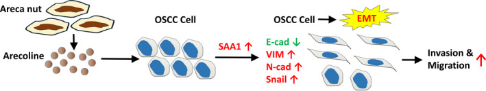
Arecoline induces EMT and promotes metastasis of oral cancer by upregulation of SAA1. As shown in the graph, arecoline induces EMT and promotes metastasis through regulation of the secretion of inflammatory markers, especially the secretion of SAA1 in OSCC
CONFLICT OF INTEREST
The authors have no conflict of interest.
Supporting information
Fig S1
Fig S2
Fig S3
Fig S4
Fig S5
Table S1
"V体育官网入口" ACKNOWLEDGMENTS
This work was supported by the Guangdong Basic and Applied Basic Research Foundation (2020A1515010291), and Youth Program of National Natural Science Foundation of China (82002871).
Ren H, He G, Lu Z, et al. Arecoline induces epithelial‐mesenchymal transformation and promotes metastasis of oral cancer by SAA1 expression. Cancer Sci. 2021;112:2173–2184. 10.1111/cas.14866
REFERENCES
- 1. Lee CC, Hsiao CY, Lee SC, et al. Suppression of oral cancer by induction of cell cycle arrest and apoptosis using Juniperus communis extract. Biosci Rep. 2020;40(9):BSR20202083. [DOI] [PMC free article] [PubMed] [Google Scholar]
- 2. Kumar M, Nanavati R, Modi TG, et al. Oral cancer: etiology and risk factors: a review. J Cancer Res Ther. 2016;12(2):458‐463. [DOI] [PubMed] [Google Scholar]
- 3. Chen WQ, Zheng RS, Baade PD, et al. Cancer statistics in China, 2015. CA: A Cancer J Clin, 2016;66(2):115‐132. [DOI] [PubMed] [Google Scholar]
- 4. Siegel RL, Miller KD, Jemal A, et al. Cancer statistics, 2019. CA A Cancer J Clin. 2019;69(1):7‐34. [DOI] [PubMed] [Google Scholar]
- 5. Islam S, Muthumala M, Matsuoka H, et al. How each component of betel quid is involved in oral carcinogenesis: mutual interactions and synergistic effects with other carcinogens–‐a review article. Curr Oncol Rep. 2019;21:53. [DOI] [PubMed] [Google Scholar]
- 6. Islam S, Uehara O, Matsuoka H, et al. DNA hypermethylation of sirtuin 1 (SIRT1) caused by betel quid chewing‐a possible predictive biomarker for malignant transformation. Clin Epigenetics. 2020;12(1):12. [DOI (V体育平台登录)] [PMC free article] [PubMed] [Google Scholar]
- 7. Wang H, Deng X, Zhang J, et al. Elevated expression of zinc finger protein 703 promotes cell proliferation and metastasis through PI3K/AKT/GSK‐3beta signaling in oral squamous cell carcinoma. Cell Physiol Biochem. 2017;44:920‐934. [DOI] [PubMed] [Google Scholar]
- 8. Xu Z, Huang CM, Shao Z, et al. Autophagy induced by areca nut extract contributes to decreasing cisplatin toxicity in oral squamous cell carcinoma cells: roles of reactive oxygen species/AMPK signaling. Int J Mol Sci. 2017;18(3):524. [DOI] [PMC free article] [PubMed] [Google Scholar]
- 9. Illeperuma RP, Kim DK, Park YJ, et al. Areca nut exposure increases secretion of tumor‐promoting cytokines in gingival fibroblasts that trigger DNA damage in oral keratinocytes. Int J Cancer. 2015;137:2545‐2557. [DOI] [PMC free article] [PubMed] [Google Scholar]
- 10. Chang MC, Chen YJ, Chang HH, et al. Areca nut components affect COX‐2, cyclin B1/cdc25C and keratin expression, PGE2 production in keratinocyte is related to reactive oxygen species, CYP1A1, Src, EGFR and Ras signaling. PLoS One. 2014;9:e101959. [DOI] [PMC free article] [PubMed] [Google Scholar]
- 11. Hsieh YP, Wu KJ, Chen HM, et al. Arecoline activates latent transforming growth factor beta1 via mitochondrial reactive oxygen species in buccal fibroblasts: suppression by epigallocatechin‐3‐gallate. J Formos Med Assoc. 2018;117:527‐534. [DOI] [PubMed] [Google Scholar]
- 12. Asthana S, Labani S, Kailash U, et al. Association of smokeless tobacco use and oral cancer: a systematic global review and meta‐analysis. Nicotine Tob Res. 2019;21(9):1162‐1171. [DOI (VSports app下载)] [PubMed] [Google Scholar]
- 13. Adhikari BR, Yoshida K, Paudel D, et al. Aberrant expression of DUSP4 is a specific phenomenon in betel quid‐related oral cancer. Med Mol Morphol. 2020. 10.1007/s00795-020-00265-3 [V体育2025版 - DOI] [PubMed] [Google Scholar]
- 14. Hsieh YH, Syu RJ, Lee CC, et al. Arecoline induces epithelial mesenchymal transition in HK2 cells by upregulating the ERK‐mediated signaling pathway. Environ Toxicol. 2020;35(9):1007‐1014. [DOI] [PubMed] [Google Scholar]
- 15. Fang C‐Y, Hsia S‐M, Hsieh P‐L, et al. Slug mediates myofibroblastic differentiation to promote fibrogenesis in buccal mucosa. J Cell Physiol. 2019;234(5):6721‐6730. [DOI] [PubMed] [Google Scholar]
- 16. Lu Z, He Q, Liang J, et al. miR‐31‐5p is a potential circulating biomarker and therapeutic target for oral cancer. Mol Ther Nucleic Acids. 2019;7(16):471‐480. [DOI] [PMC free article] [PubMed] [Google Scholar]
- 17. Chen P‐H, Mahmood Q, Mariottini GL, et al. Adverse health effects of betel quid and the risk of oral and pharyngeal cancers. Biomed Res Int. 2017;2017:3904098. [DOI] [PMC free article] [PubMed] [Google Scholar]
- 18. Shih Y‐H, Chiu K‐C, Wang T‐H, et al. Effects of melatonin to arecoline‐induced reactive oxygen species production and DNA damage in oral squamous cell carcinoma. J Formos Med Assoc. 2021;120(1 Pt 3):668‐678. [DOI] [PubMed] [Google Scholar]
- 19. Shiah S‐G, Hsiao J‐R, Chang W‐M, et al. Downregulated miR329 and miR410 promote the proliferation and invasion of oral squamous cell carcinoma by targeting Wnt‐7b. Cancer Res. 2014;74(24):7560‐7572. [VSports注册入口 - DOI] [PubMed] [Google Scholar]
- 20. Zhong X, Lu Q, Zhang QI, et al. Oral microbiota alteration associated with oral cancer and areca chewing. Oral Dis. 2021;27(2):226‐239. [DOI] [PubMed] [Google Scholar]
- 21. Pastushenko I, Blanpain C. EMT transition states during tumor progression and metastasis. Trends Cell Biol. 2019;29(3):212‐226. [DOI] [PubMed] [Google Scholar]
- 22. Mittal V. Epithelial mesenchymal transition in tumor metastasis. Annu Rev Pathol. 2018;24(13):395‐412. [DOI] [PubMed] [Google Scholar]
- 23. Kim S‐E, Shin S‐H, Lee J‐Y, et al. Transition states during tumor progression and metastasis. Resveratrol induces mitochondrial apoptosis and inhibits epithelial‐Mesenchymal transition in oral squamous cell carcinoma cells. Nutr Cancer. 2018;70:125‐135. [DOI] [PubMed] [Google Scholar]
- 24. Ho CM, Hu F‐W, Lee S‐S, et al. ZEB1 as an indicator of tumor recurrence for areca quid chewing‐associated oral squamous cell carcinomas. J Oral Pathol Med. 2015;44:693‐698. [DOI (VSports手机版)] [PubMed] [Google Scholar]
- 25. Zheng L, Jian X, Guo F, et al. miR‐203 inhibits arecoline‐induced epithelial‐mesenchymal transition by regulating secreted frizzled‐related protein 4 and transmembrane‐4 L six family member 1 in oral submucous fibrosis. Oncol Rep. 2015;33(6):2753‐2760. [DOI] [PubMed] [Google Scholar]
- 26. Peng C‐Y, Liao Y‐W, Lu M‐Y, et al. Positive feedback loop of SNAIL‐IL‐6 mediates myofibroblastic differentiation activity in precancerous oral submucous fibrosis. Cancers (Basel). 2020;12(6):1611. [DOI] [PMC free article] [PubMed] [Google Scholar]
- 27. Zheng L, Guan Z‐J, Pan W‐T, et al. Tanshinone suppresses arecoline‐induced epithelial‐mesenchymal transition in oral submucous fibrosis by epigenetically reactivating the p53 pathway. Oncol Res. 2018;26(3):483‐494. [VSports - DOI] [PMC free article] [PubMed] [Google Scholar]
- 28. Chang LY, Wan HC, Lai YL, et al. Areca nut extracts increased the expression of cyclooxygenase‐2, prostaglandin E2 and interleukin‐1alpha in human immune cells via oxidative stress. Arch Oral Biol. 2013;58:1523‐1531. [DOI] [PubMed] [Google Scholar]
- 29. Sun L, Ye RD. Serum amyloid A1: structure, function and gene polymorphism. Gene. 2016;583(1):48‐57. [DOI] [PMC free article] [PubMed] [Google Scholar]
- 30. Yang MU, Liu F, Higuchi K, et al. Serum amyloid a expression in the breast cancer tissue is associated with poor prognosis. Oncotarget. 2016;7:35843‐35852. [DOI] [PMC free article] [PubMed] [Google Scholar]
- 31. Apanovich N, Peters M, Apanovich P, et al. The genes‐candidates for prognostic markers of metastasis by expression level in clear cell renal cell cancer. Diagnostics (Basel). 2020;10(1):30. [DOI] [PMC free article] [PubMed] [Google Scholar]
- 32. Wu D‐C, Wang K‐Y, Wang SSW, et al. Exploring the expression bar code of SAA variants for gastric cancer detection. Proteomics. 2017;17(11). ["V体育2025版" DOI] [PubMed] [Google Scholar]
- 33. Zhang Y, Wei Y, Jiang B, et al. Scavenger receptor A1 prevents metastasis of non‐small cell lung cancer via suppression of macrophage serum amyloid A1. Cancer Res. 2017;77(7):1586‐1598. [DOI] [PubMed] [Google Scholar]
- 34. Takehara M, Sato Y, Kimura T, et al. Cancer‐associated adipocytes promote pancreatic cancer progression through SAA1 expression. Cancer Sci. 2020;111(8):2883‐2894. [DOI] [PMC free article] [PubMed] [Google Scholar]
- 35. Li ZE, Hou Y, Zhao M, et al. Serum amyloid a, a potential biomarker both in serum and tissue, correlates with ovarian cancer progression. J Ovarian Res. 2020;13(1):67. [DOI] [PMC free article] [PubMed] [Google Scholar]
- 36. Chang P‐Y, Kuo Y‐B, Wu T‐L, et al. Association and prognostic value of serum inflammation markers in patients with leukoplakia and oral cavity cancer. Clin Chem Lab Med. 2013;51(6):1291‐1300. ["V体育平台登录" DOI] [PubMed] [Google Scholar]
- 37. Arun I, Maity N, Hameed S, et al. Lymph node characteristics and their prognostic significance in oral squamous cell carcinoma. Head Neck. 2021;43(2):520‐533. [DOI] [PubMed] [Google Scholar]
- 38. Li B, Hou DQ, Xu SB, et al. TLR2 deficiency enhances susceptibility to oral carcinogenesis by promoting an inflammatory environment. Am J Cancer Res. 2019;9(12):2599‐2617. [PMC free article] [PubMed] [Google Scholar]
- 39. Wu T‐F, Chen L, Bu L‐L, et al. CD44+ cancer cell‐induced metastasis: a feasible neck metastasis model. Eur J Pharm Sci. 2017;101:243‐250. [DOI] [PubMed] [Google Scholar]
Associated Data
This section collects any data citations, data availability statements, or supplementary materials included in this article.
"VSports app下载" Supplementary Materials
Fig S1
Fig S2
Fig S3
Fig S4
Fig S5
Table S1